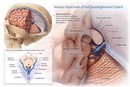
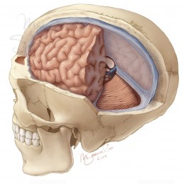
This neuroanatomical illustration explains the complex venous pathways that travel through the quadrigeminal cistern, a dilation of subarachnoid space between the midbrain, cerebral hemispheres, and cerebellum.
To accurately depict the intricate neuroanatomy in this region, I reconstructed anonymized patient CT and MRI scans to create 3D models that I referenced for accurate size and structure relationships.
Continue reading below to learn more about how I created these reference models from DICOM data, and how I translated these models to a rendered illustration.
Segmenting DICOM data in OsiriX and importing to Zbrush
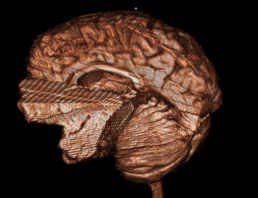
Using the DICOM segmentation features of OsiriX software, I isolated the soft tissue structures of the brain from an anonymized MRI scan and the skull from a CT scan of another patient. I generated these data sets as 3D models using the 16-bit threshold editor and explored the anatomy with various slices through the models (below).
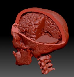
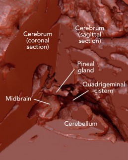
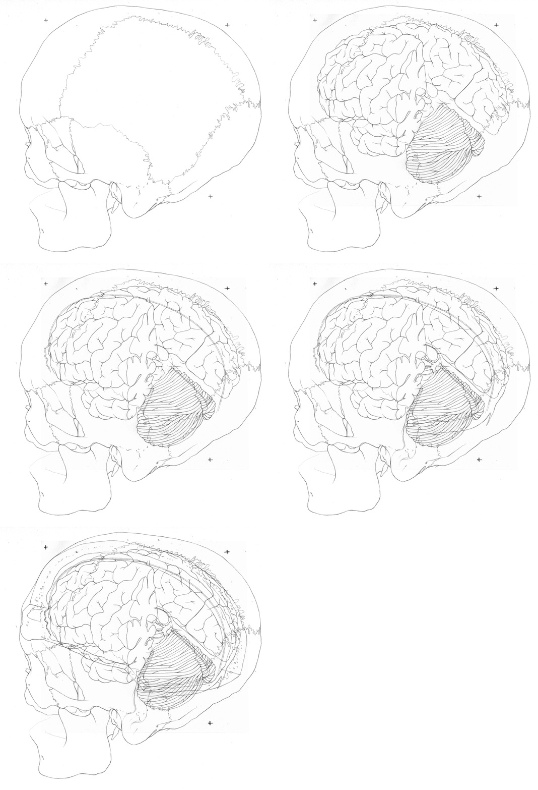
After exporting the whole models and importing them to Zbrush, I aligned them as if they had existed in the same patient. I then used a live Boolean subtraction to create a clean window in the models and visualize the internal structures of the quadrigeminal cistern.
Beginning the Illustration
Referencing my combined model in an oblique view as well as the original 3D reconstructed patient models and neuroanatomy reference books, I created a clean outline to preserve anatomical relationships and detail in my final illustration. This was achieved by creating separate traditional pencil outlines for each structure (skull, cerebrum & cerebellum, cut edge of the dura, venous structures, and the cut edge of the cranium) and aligning them in Adobe Photoshop.
